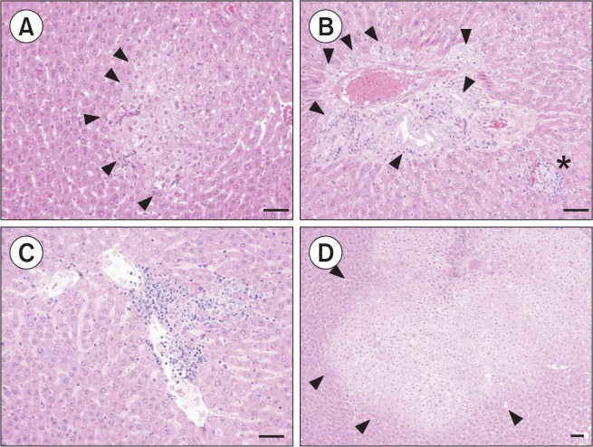
Fig. 2.
Histopatholgical findings in the liver of BPA-treated juvenile rats. BPA 0.5 mg/kg: Photomicrograph of the eosinophilic altered cell foci (arrow head) with grade + (A), BPA 0.5 mg/kg: Photomicrograph of minimal bile duct hyperplasia (arrow head) with lymphocytic cell infiltration (asterisk) with grade ++ (B). BPA 0.5 mg/kg: Focal inflammatory cell infiltration with grade + (C). BPA 5 mg/kg: Photomicrograph of the eosinophilic altered cell foci (arrow head) with grade ++ (D). (Bar=50 μm).

