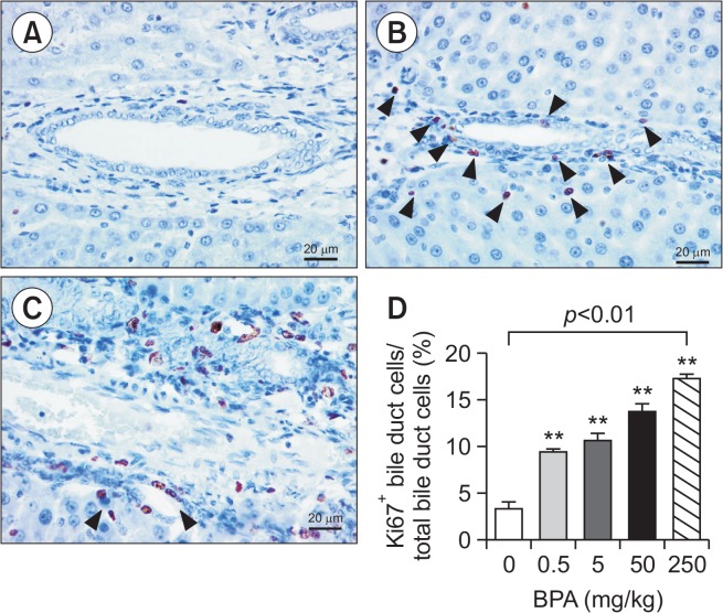
Fig. 3.
Bile duct proliferation in the liver of BPA-treated juvenile rats. Photomicrograph of the immunohistochemistry for Ki-67 to evaluate bile duct proliferation, BPA 0 mg/kg (A), 0.5 mg/kg with arrow heads indicating Ki-67+ cells (B) and 250 mg/kg (C). Ratio of Ki-67+ bile duct cells over total bile duct cells (mean ± SEM of counting in 7–45 sections). **p<0.01 One-way ANOVA followed by Dunnett’s post hoc analysis.

