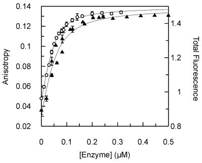
Figure 4.
Binding of D88N mutant UDG to 2-aminopurine and HEX-labeled oligonucleotides. Equilibrium binding experiments were performed with the HEX-labeled oligonucleotide 1HU and the 2-aminopurine-containing oligonucleotide 1U (Materials and Methods). Fluorescence anisotropy (left scale) was used to follow the binding of D88N to 1HU (open circles; Figure 2), while total 2-aminopurine fluorescence intensity (right scale) was used to measure binding to 1U (closed triangles). Both data sets are shown with the best fit to equation 1, with the following values: 1HU, Kd = 0.012 ± 0.001 µM, AD = 0.042 ± 0.001, ADE = 0.140 ± 0.001; 1U, Kd = 0.024 ± 0.006 µM, FD = 0.86 ± 0.03, FDE = 1.49 ± 0.02 (where FD and FDE are the total fluorescence of free and enzyme-bound DNA, respectively)

