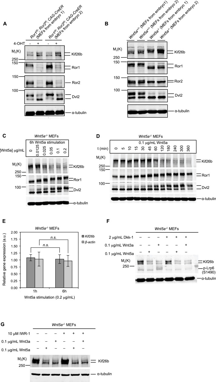
Figure 2. Kif26b protein levels are regulated by the expression of Wnt5a and Ror proteins in primary mouse embryonic fibroblasts.
(A) Immunoblots of Kif26b protein, Ror1 protein, Ror2 protein and Dvl2 protein in primary MEFs derived from two E12.5 Ror1f/f; Ror2f/f; CAG-CreER mice. 4-OHT or vehicle control was added for 72 hr prior to lysis. Results of two independent biological replicates are shown. (B) Immunoblots of Kif26b protein, Ror1 protein, Ror2 protein and Dvl2 protein in primary MEFs derived from littermate E12.5 Wnt5a+/+ and Wnt5a-/- mice. Results of two independent biological replicates are shown. (C) Immunoblots of Kif26b protein, Ror1 protein and Dvl2 protein in primary MEFs derived from E12.5 Wnt5a-/- mice. Recombinant Wnt5a protein was added for 6 hr prior to lysis at the indicated dose. (D) Immunoblots of Kif26b protein, Ror1 protein and Dvl2 protein in primary MEFs derived from E12.5 Wnt5a-/- mice. 0.1 μg/ml recombinant Wnt5a protein was added for the indicated amount of time prior to lysis. (E) Plot showing relative mRNA expression of Kif26b and β-actin as measured by RT-qPCR in primary MEFs derived from E12.5 Wnt5a-/- mice, with 1 hr or 6 hr of Wnt5a stimulation. The y-axis represents fold change relative to expression levels in unstimulated cells. Error bars represent ± SEM calculated from three technical replicates. t-test (unpaired) was determined for the following comparisons: Kif26b 1 hr vs. 6 hr, p=0.249, not significant; β-actin 1 hr vs. 6 hr, p=0.320, not significant. (F) Immunoblots of Kif26b protein and phospho-Lrp6 (serine 1490) protein in primary MEFs derived from E12.5 Wnt5a-/- mice. Recombinant Dkk-1 (0.1 μg/ml) or vehicle control was added 8 hr prior to lysis. Recombinant Wnt3a protein, Wnt5a protein or vehicle control was added for 6 hr prior to lysis. (G) Immunoblots of Kif26b protein in primary MEFs derived from E12.5 Wnt5a-/- mice. IWR-1 or vehicle control was added 7 hr prior to lysis. Recombinant Wnt3a protein, Wnt5a protein or vehicle control was added for 6 hr prior to lysis. α-tubulin was used for loading controls in all experiments. All immunoblot samples were normalized by BCA assays for total protein.


