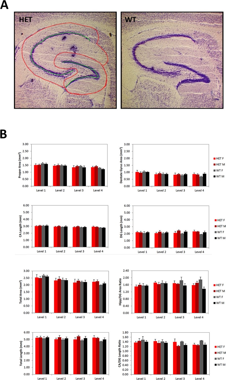
Fig 3. Hippocampal size does not change in Slc1a1-HET mice.

Representative images (A) and morphometric analysis (B) of hippocampus in Nissl-stained sagittal brain sections of Slc1a1-HET (n = 8) and WT (n = 5) mice. A) The total area of the hippocampal formation, the subareas of the hippocampus proper and DG, and the ratio of the hippocampus proper area to the DG area, as well as the length of the stratum pyramidale of the complete CA, the length of the stratum granulosum in the DG, the total length of these combined, and the ratio of CA to DG lengths were measured. These measurements were obtained on 4 section levels through each hippocampus of each animal. B) A two-way (Sex x Genotype) repeated measures ANOVA combining all available measurements for each of the section levels for each animal was performed. WT, wildtype; HET, Slc1a1-heterozygous. DG, dentate gyrus; CA, cornu ammonis.
