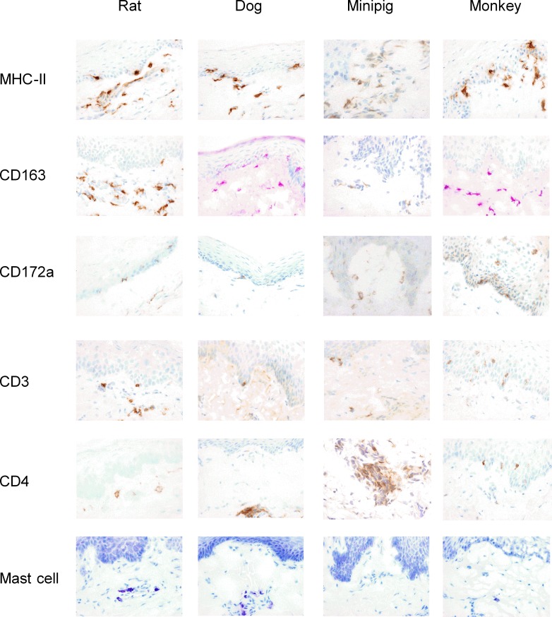
Fig 4. Mapping of immune cells in the ventral surface of tongues from rats, dogs, minipigs and monkeys.
Mucosal tissues from animal species were processed for immunohistology analysis, as described in Methods. Slides were stained with the following specific antibodies: anti-MHC-II, anti-CD163, anti-CD172a, anti-CD3, and anti-CD4 to detect and quantify positive cells in tissue sections or with toluidine blue to quantify mast cells (magnification x200). Representative photomicrographs of mucosal tissue sections are shown.

