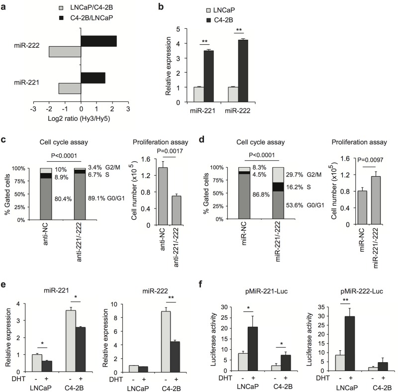
Fig 1. Overexpression of miR-221/-222 promotes CRPC cell proliferation.
(a) MiRNA microarray results showing upregulation of miR-221/-222 in C4-2B cells compared to LNCaP cells. (b) RT-qPCR confirmed microarray results. The expression level was normalized by U6 snRNA. (c) C4-2B cells were co-transfected with miR-221 and miR-222 inhibitors (anti-221/-222) and non-specific control (NC). Cell number was counted 3 days after transfection. Cell cycle analysis was performed in parallel. (d) C4-2B cells were co-transfected with miR-221 and miR-222 precursors (miR-221/-222) and analyzed as described in (c). (e). LNCaP and C4-2B cells were grown in phenol red-free RPMI 1640 media containing 5% charcoal-stripped fetal bovine serum (CSS) for 3 days followed by treatment with 10 nM dihydrotestosterone (DHT) or ethanol control for 16 hours. MiR-221/-222 were examined using RT-qPCR. (f) LNCaP and C4-2B cells were grown in phenol red-free RPMI 1640 media containing 5% CSS with or without 10 nM DHT for 2 days. pMiR-221-Luc or pMiR-222-Luc luciferase constructs containing DNA sequences at 3’UTR complementary to miR-221/-222 were transfected into cells. pRL-TK Renilla luciferase reporter was co-transfected as an internal control. Luciferase activity (Firefly/Renilla ratio) was determined 24 hours after transfection. The p-value for cell cycle distribution of 10,000 gated cells was determined using a chi-squared test. The p-value for other assays was determined using a two-tailed Student’s t-test. Data presented are mean ± SD of three measurements. * P < 0.05; ** P < 0.01.

