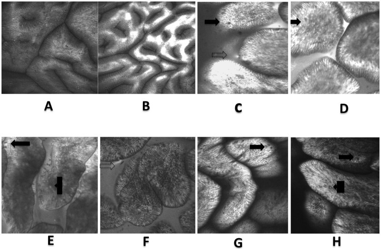
Fig 2.
Confocal laser endomicroscopy images: A- normal antrum, B- normal antrum showing confinement of fluorescein within the mucosa. C to E shows CLE images of gastric intestinal metaplasia and F to H: comparative CLE images of the duodenum. Visible in both sets are the presence of villus-like structures, goblet cells (solid arrows), intraepithelial lymphocytes (box arrows), plumes (dotted arrows) and a visible brush borders.

