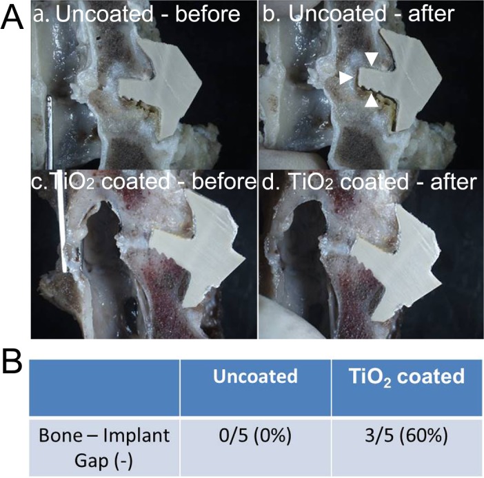
Fig 4.

(A) The μ-CT images. (a) Uncoated PEEK. Bone cysts and osteolytic lesion were observed surrounding the PEEK implant (black arrowheads). (b) TiO2-coated PEEK. An intervertebral bony bridge was observed (white arrowheads). (B) The bony union rate of the PEEK implants based on the μ-CT finding.
