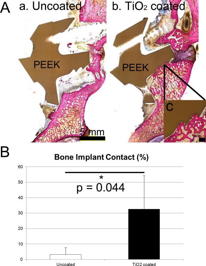
Fig 6.
(A) Histology. (a) Uncoated PEEK. Osteolytic lesion was observed surrounding the PEEK implant. (b) TiO2-coated PEEK. Bone–implant integration was observed on the lower surface of the PEEK implant. Another section of this specimen showed direct apposition on the upper surface, but not on the lower surface. This specimen was considered to be fused because there was no gap during the manual palpation analysis. (c) Magnified view. (B) The bone–implant contact ratio based on histomorphometry.

