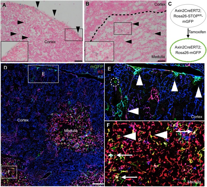
Fig 1. Postnatal thymic epithelium contains Axin2-expressing cells in both cortex and medulla.
(A to B) P16 Axin2-lacZ thymus stained with X-gal. Arrowheads point to X-gal stained cells, and insets are magnified views of the boxed regions. (C to F) AXCT2;mTmG thymus traced with a high dose of tamoxifen (15mg tamoxifen/25g body weight) from P16-P18 and stained for K5 and K8 expression. Boxed regions in (D) are magnified in (E) and (F). (E) Cortical region showing clusters of AXCT2-labeled K8+ cTECs after two days (arrowhead). Some cells with circular morphology are observed, and are absent from the thymus after one month. (F) Medullary region showing a more dispersed pattern of AXCT2-labeled cells, which express K5 only (arrowhead), or both K5 and K8 (arrow). Scale bars for (A) and (B) represent 40μm, and 100μm for (D).

