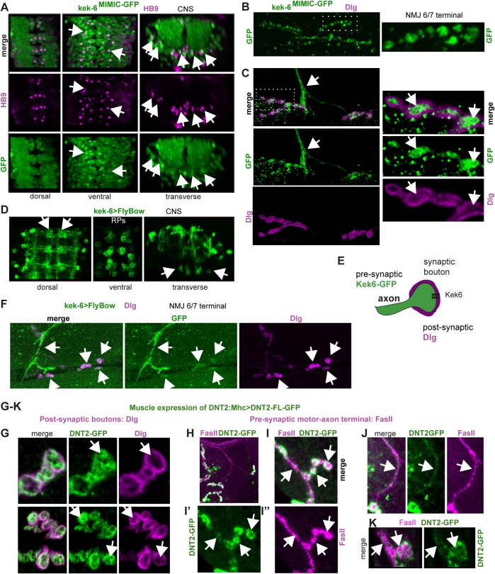
Fig 3. Kek-6 is expressed pre-synaptically in motoneurons and binds post-synaptic DNT2.
(A) In Kek-6GFP larval VNCs, GFP colocalises with the neuronal marker HB9 (arrows show examples). (B) Kek-6GFP was found in third instar larval muscle 6/7 NMJ and synaptic boutons (dotted rectangle: higher magnification, right). (C) Kek-6GFP was found in the motoneuron axonal terminal (arrows), and in pre-synaptic bouton lumen (dotted rectangle: higher magnification, right), not colocalising with the post-synaptic marker anti-Dlg (arrows).(D) Kek-6>FlyBow was localized to CNS axons and dendrites (arrows), and cell bodies of the RP3,4,5 motoneuron clusters (ventral and transverse views, arrows). (E) Illustration. (F) Kek-6>FlyBow was also distributed along the motoneuron axons, NMJ terminal (arrow) and synaptic boutons (arrows). (G-K) Over-expression of GFP tagged full-length DNT2 in muscle (MhcGAL4>UAS-DNT2-FL-GFP) revealed: (G) DNT2-GFP distribution within the pre-synaptic bouton lumen (arrows), boutons labeled post-synaptically with anti-Dlg; (H-K) DNT2-GFP along the motoraxon (labeled with anti-FasII) and within the pre-synaptic bouton lumen (arrows).

