
Figure 2. Relationships between pericardial fat and left ventricular geometry and function.
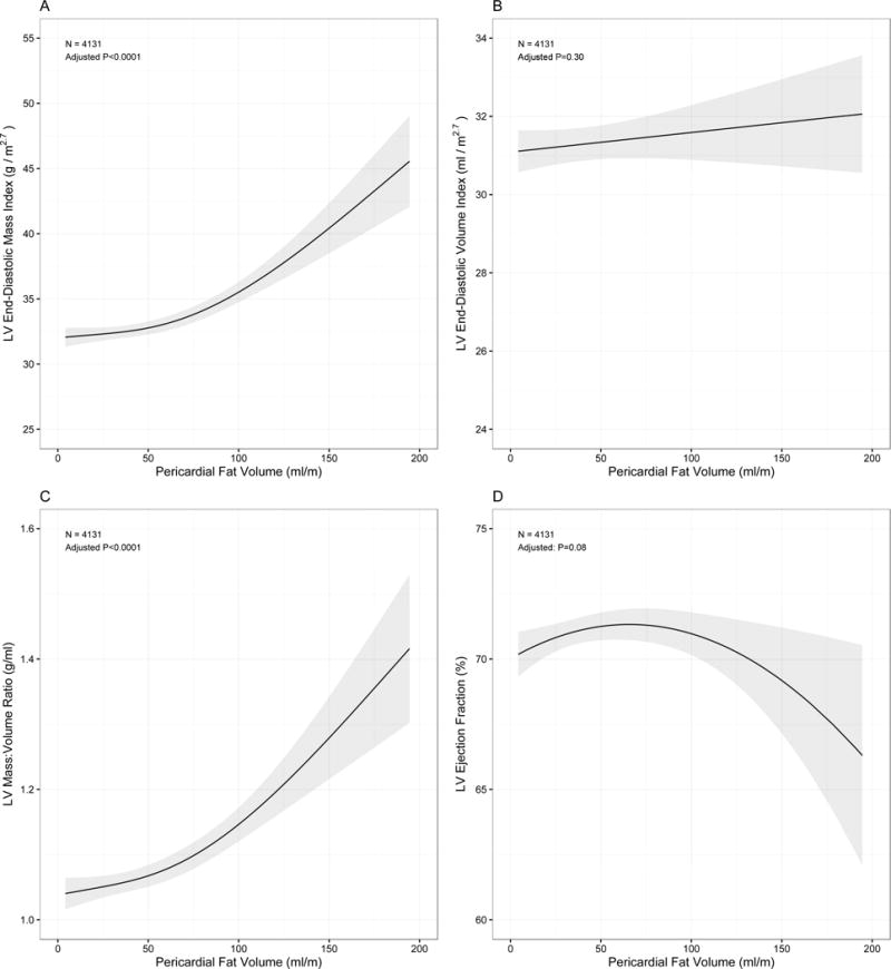
Associations for LV mass (panel A), LV mass-to-volume ratio (panel C) and ejection fraction were curvilinear (panel D), with significant quadratic terms (Table 4). There was no significant relationship noted between pericardial fat and LV volume (panel B). Graphs and p-values are derived from fully adjusted generalized additive spline models with grey bands represent 95% confidence intervals.