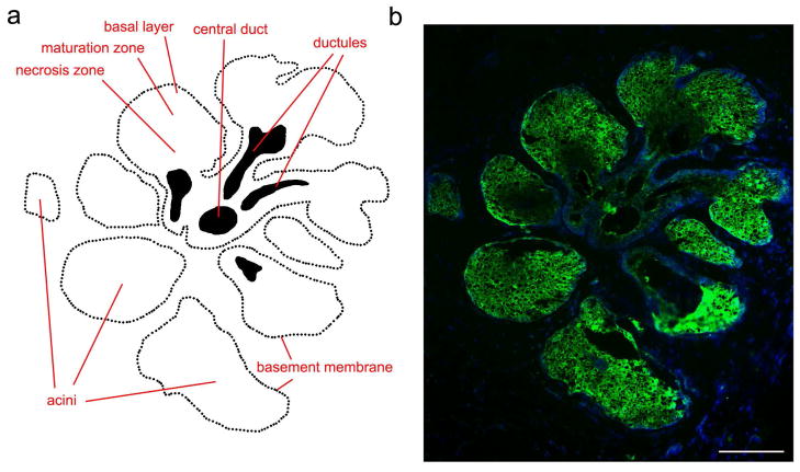
Figure 1. Proteolytic activity in the normal human meibomian gland.
(a) Schematic representation of the structure of the meibomian gland. Cells at varying degrees of differentiation are present within the secretory acini, corresponding to proliferating cells in the basal layer and differentiating cells that accumulate intracellular lipid droplets in the maturation zone. Fully differentiated cells are present in the necrosis zone, where the meibum is produced and then released through small ductules into a long central duct. (b) In situ gelatin zymography (green) showing localization of gelatinolytic activity in secretory acini of a meibomian gland, shown schematically in (a). Within the acinus, activity was detected in proliferating and differentiating cells, and decreased towards the necrosis zone. Nuclei were counterstained using DAPI (blue). Scale bar, 100 μm. Adapted from Mauris et al. (Mauris et al., 2015).

