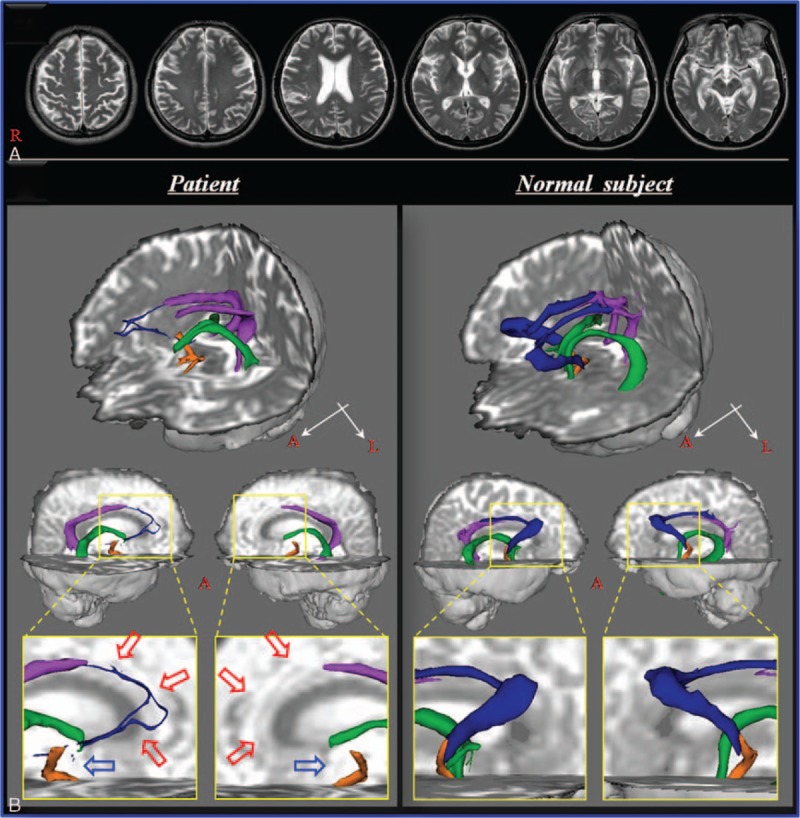
Figure 1.

(A) T2-weighted brain MR images at 10 weeks after onset show no abnormality. (B) Results of diffusion tensor tractography of the Papez circuit. On 10-week diffusion tensor tractography, discontinuation of the fornical column is observed in both hemispheres (blue arrows), and thinning of the thalamocingulate tract is observed in the right hemisphere and nonreconstruction in the left hemisphere (red arrows). MR = magnetic resonance.
