
Figure 2.
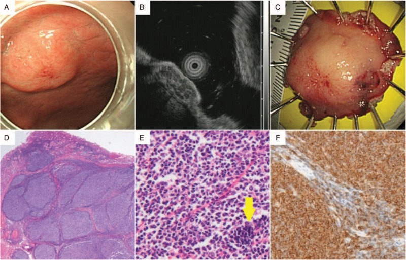
A lesion showing the histology of follicular lymphoma after endoscopic submucosal dissection (ESD). (A) Upper endoscopy showed a well defined, polypoid gastric subepithelial lesion with a size of about 1.5 cm in the middle body, anterior wall. This lesion had a central mucosal erythema that is believed to be a scar created by a previous biopsy that was performed at another medical center. (B) On endoscopic ultrasonography, the lesion appeared as a 15 mm × 5 mm hypoechoic mass in the submucosa layer without infiltration of the muscularis propria. It was presumed to be a neuroendocrine tumor, even though the lesion had inhomogeneous and multiseptated morphological features. (C) The ESD specimen of this lesion appeared to be 2.8 × 2.4 × 0.6 cm3 in size after ESD, which had been performed without complications. (D) Histopathological analysis of the ESD specimen showed lymphoid hyperplasia below the normal gastric mucosa (hematoxylin and eosin stain, original magnification ×12). (E) This structure showed back-to-back arrangement and a very thin mantle zone (hematoxylin and eosin, original magnification ×200). (F) Lymphocytes of the abnormal structure were positive for CD20; this finding was compatible with follicular lymphoma (hematoxylin and eosin, original magnification ×200).