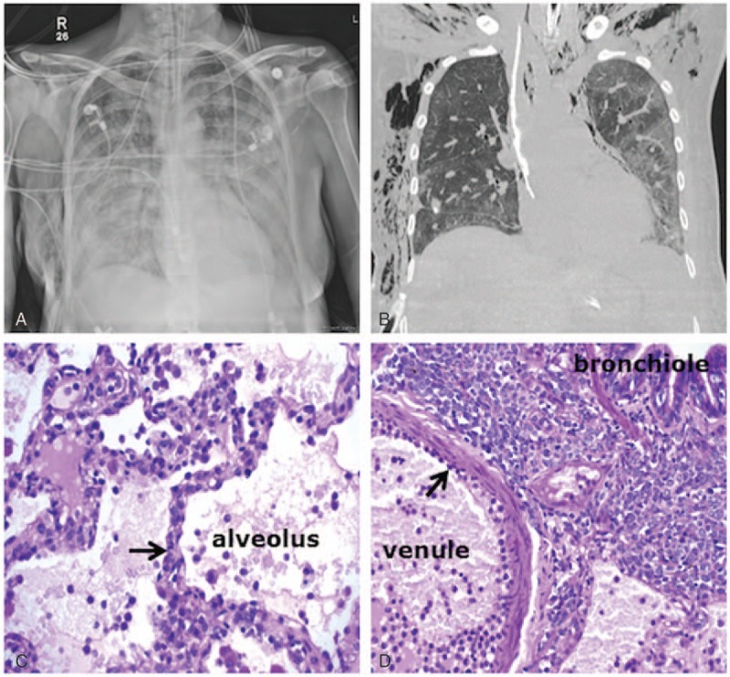
Figure 4.

Radiological–pathological correlation in a 55-year-old female patient diagnosed with monocytic AML and presenting with hyperleukocytosis (WBC = 254 × 109/L) and pulmonary leukostasis with acute hypoxic respiratory failure requiring mechanical ventilation. Initial chest radiograph showed diffuse, bilateral airspace and interstitial opacities (A). The patient underwent open lung biopsy and a CT scan obtained at day 5 for air leakage showed persistent diffuse ground-glass opacities, more confluent on the left side and dependently (B). Bottom panels: periodic acid–Schiff stain of the lung biopsy. (C) Leukostasis with capillary obstruction by leukemic cells (blue stain, black arrow) within the alveolar–capillary barrier. (D) Leukemic cells adhering to the endothelium (black arrow) and massively infiltrating the interstitium between the venule and the bronchiole. AML = acute myeloid leukemia, CT = computed tomography, WBC = white blood cell.
