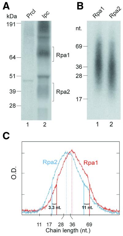
Figure 2.

Sizing nascent SV40 DNA chains radiolabeled in the DNA moiety and crosslinked to RPA subunits. (A) Photolabeled RPA was isolated from the pulse-labeled replication mixture without DNase I treatment, as detailed in Figure 1 and Materials and Methods. Precipitates of pre-clearing (lane 1) and IP of RPA under native conditions (lane 2) were resolved by SDS–PAGE. (B) The Rpa1 and Rpa2 bands were excised from the indicated gel sections. The photolabeled proteins were extracted and treated with proteinase K. After phenol extraction, the released nascent DNA was separated by electrophoresis on 15% TBU gel (Novex). (C) Densitometric profiles of (B), lane 1 (red) and lane 2 (cyan). Peak values and half peak values marking distributions borders are indicated by respective full or dashed vertical lines in corresponding color. kDa, protein size markers; nt, nucleotide size markers; Prcl, pre-clearing immunoprecipitate; Ipc, specific immunoprecipitate.
