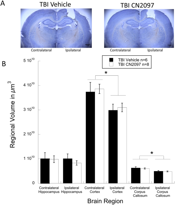
Figure 7.

Brain regional volume analysis in CN2097- versus vehicle-treated rats. Photomicrographs (A) show coronal brain sections from ipsilateral and contralateral hemispheres from vehicle- and CN2097-treated rats, demonstrating comparable injury profiles. Bar graph (B) shows regional brain volume measurements for vehicle-treated (TBI Vehicle; n = 6) and CN2097-treated (TBI CN2097; n = 8) rats, indicating comparable ipsilateral and contralateral regional brain volumes of the cerebral cortex, hippocampus, and corpus callosum. *Indicates significant (p < 0.01) differences between ipsilateral and contralateral hemispheres for both treatment groups. Data represent mean ± SEM.
