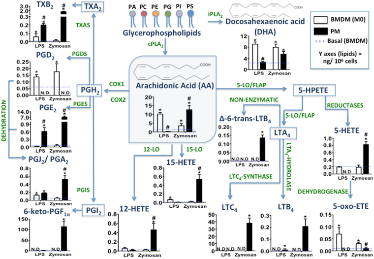
Figure 1.
Comparative eicosanoid catabolism profile in BMDMs and PMs stimulated with microbial particles. BMDMs and PMs (1 × 10⁶ cells/well) were adhered on cell culture plates for 24 h. Macrophages were stimulated with LPS (500 ng/mL) or zymosan (30 particles/cell) for 6 and 1.5 h, respectively. The lipid mediators in cell culture supernatants were identified and quantified by HPLC-MS/MS (MRM mode) for eicosanoids: TXB2, PGD2, PGE2, PGJ2/PGA2, 6-keto-PGF1α, 12-HETE, 15-HETE, 5-HETE, 5-oxo-ETE, LTC4, LTB4, and Δ-6-trans-LTB4 as well as for the release of fatty acids: AA and DHA. Results are expressed as the means ± s.e.m. of three experiments (n = 3). Differences were considered when p < 0.05, *BMDMs and PMs stimulated or not, compared to non-stimulated basal BMDM production (dashed line), #PM compared to BMDM eicosanoid production, after LPS or zymosan stimulation.

