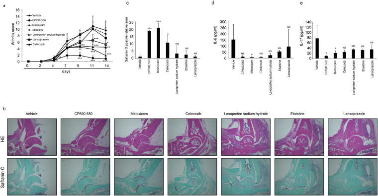
Figure 4.
Effect of Stat3 inhibitors on arthritis development in CIA models. (a–c) 5-week-old wild-type DBA/1J male mice were initially injected with type II collagen with CFA on day -21, and arthritis was induced by a second injection on day 0. Indicated drugs (each 15 mg/kg/day) were administered IP once a day for 2 weeks starting at day 0. An arthritis score was evaluated at indicated time points after the second injection (a). Histological analysis was performed using hematoxylin eosin staining (HE, upper panels) or safranin O and methyl green (lower panels) staining of ankle joints from mice treated with indicated drugs for two weeks (b), and the safranin O-positive area was quantified (c). Serum IL-6 (d) and IL-17 (e) protein levels were also examined by ELISA in these mice treated with indicated drugs for two weeks. Data represent mean arthritis score (a), IL-6 (d) or IL-17 levels (e) ± SD (a, vehicle n = 10, CP690,550 n = 8, meloxicam n = 7, celexib n = 5, ebastine, loxoprofen sodium hydrate or lansoprazole n = 4 each; d, vehicle or meloxicam n = 5 each, CP690,550, celexib, loxoprofen sodium hydrate or lansoprazole n = 4 each, ebastine n = 3; e, vehicle or CP690,550 n = 7 each, meloxicam or loxoprofen sodium hydrate n = 4 each, celecoxib, ebastine or lansoprazole n = 3 each; *P < 0.05, **P < 0.01, ***P < 0.001, NS not significant, vs vehicle). Bar, 100 µm. Data in (c) represent mean relative safranin O-positive areas ± SD (n = 3 each; *P < 0.05, ***P < 0.001, NS not significant, vs vehicle). Representatives of at least two independent experiments are shown.

