Figure 1.

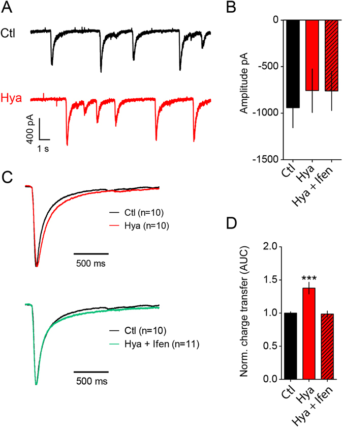
ECM removal enhances GluN2B-NMDAR mediated synaptic currents. (A) Example traces of NMDAR - mediated sEPCSs before and after Hya treatment in dissociated hippocampal cultures DIV21-24. (B) Amplitudes of single peaks show no significant differences between Hya treated or Hya plus Ifenprodil treated cultures (Ctl, −905.5 ± 179.4, n = 10; Hya, −776.2 ± 174.8, n = 10; Hya + Ifen, −758.2 ± 161.7, n = 11; average ± SEM; One-way ANOVA, P = 0.7991). (C) Average of single peaks before and after Hya treatment and after Ifenprodil application. Normalization of the amplitude illustrates the increased decay-time after Hya treatment (red line) in comparison to Ctl (black line). This can be restored after Ifenprodil application (green line). Ctl traces are identical. (D) Quantification of the area under the curve (AUC) of averaged and normalized events (left), which represent the total charge transfer revealed bigger charge transfer after ECM removal, which was reduced to control levels after blocking GluN2B-NMDAR with Ifen (Ctl, 1 ± 0.02, n = 10; Hya, 1.38 ± 0.09, n = 10; Hya + Ifenprodil, 0.98 ± 0.05, n = 11; average ± SEM; One-way ANOVA, P < 0.0001, Dunnett’s Multiple Comparison Test, ***P < 0.05).
