Figure 1.
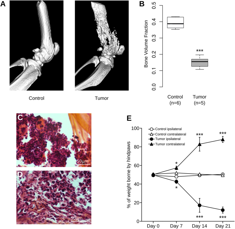
Quantification of bone destruction and assessment of nociceptive state. (A) Radiographic images of intact femur 21 days after saline (left) or sarcoma cells injection (right), revealing destruction of bone upon tumor development. (B) Bone volume quantification shows significant loss of bone density in tumor-injected group at day 21 after surgery (***P < 0.001, Mann-Whitney). (C) Hematoxylin-eosin-safran staining in control animals clearly shows normal structure of bone and bone marrow cells. (D) In contrast, in mice injected with NCTC 2472 sarcoma cells, there is a clear degradation of bone and replacement of bone marrow by sarcomatous cells. (E) Tumor-injected mice show a significant decrease in weight borne by ipsilateral paw compared to control group at days 7, 14 and 21 after surgery. Contralateral paws of tumor-injected group show a significant increase in weight borne at days 7, 14 and 21 after surgery in comparison to control group. Statistical comparison of tumor and control groups performed with two-way ANOVA with repeated measures followed by Bonferroni post-test, *P < 0.05, ***P < 0.001 compared to day 0.

