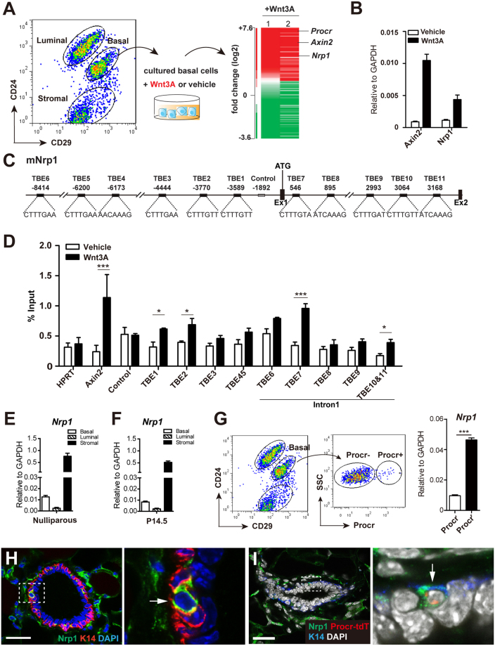
Figure 1.
Nrp1 is upregulated by Wnt signaling in MaSCs. (A) Mammary basal cells were FACS-sorted from 8-week-old nulliparous mammary gland and cultured in 3D Matrigel in the presence of Wnt3A protein or vehicle. Microarray analysis of the cultured cells indicated that Nrp1 was upregulated with Wnt3A treatment. 1 and 2 represented two independent experiments. (B) qPCR analysis validating the increased expression of Nrp1 in Wnt3A treated cells. Axin2 serves as a positive control. (C) Schematic illustration of the promoter and first intron of mouse Nrp1. TCF-binding elements (TBEs) and negative control region are indicated. Ex, Exon; Int, Intron. (D) ChIP-qPCR analysis with anti-β-catenin antibody. qPCR results were normalized to HPRT. Axin2 promoter region with known effective TEB serves as positive control. (E,F) qPCR analysis of nulliparous mammary gland (E) and pregnant day 14.5 (P14.5) (F) indicating that Nrp1 expression is higher in basal cells compared to luminal cells, and it has the highest expression in stromal cells. (G) qPCR analysis of FACS-isolated Procr+ and Procr− basal cells from nulliparous mammary gland indicating that Nrp1 is mainly expressed in Procr+ basal cells. Data are pooled from three independent experiments, are presented as mean ± SEM. ***P < 0.001. (H) Immunohistochemistry indicating that Nrp1 is expressed in a subpopulation of basal cells (arrow). Basal cells are marked by the expression of Keratin 14 (K14) in red. (I) Immunohistochemistry indicating Nrp1 expression (green) in a tdTomato+ Procr-expressing cell (Procr-tdT). Scale bar, 20 μm.

