Figure 3.
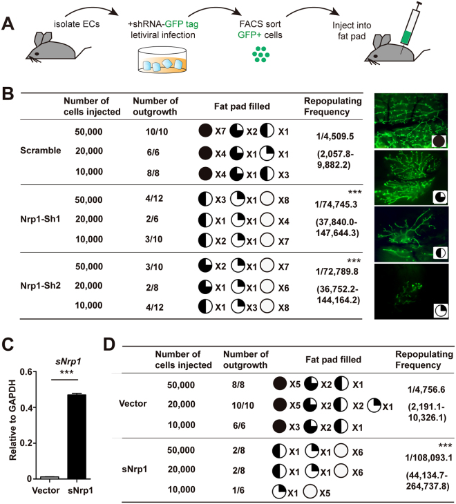
Inhibition of Nrp1 attenuates MaSCs’ reconstitution ability in vivo. (A) Schematic illustration of transplantation assay setup. Mammary epithelial cells (ECs) were isolated from 8-week-old nulliparous mice, followed by infection with scramble or Nrp1 shRNAs lentivirus. After 6 days of culture, positively infected cells (GFP+) were FACS isolated and transplanted into the cleared fat pad of 3-week-old nude mice and allowed for the formation of mammary outgrowth. (B) 10,000 or 20,000 or 50,000 of infected cells were injected into each of the cleared fat pad, and the mammary outgrowth were examined at 8 weeks post transplantation. Number of outgrowths and percentage of fat pad filled are shown. Representative images of different percentage of fat pad filled are shown on the right panels. Data are pooled from three independent experiments. (C) qPCR analysis of soluble Nrp1 (sNrp1) expression in infected (GFP+) epithelial cells. (D) 10,000 or 20,000 or 50,000 of infected cells with vector or sNrp1 overexpression were injected into each of the cleared fat pad, and the mammary outgrowths were examined at 8 weeks post transplantation. Data were pooled from three independent experiments. Data are presented as mean ± SEM. ***P < 0.0001.

