Figure 4.
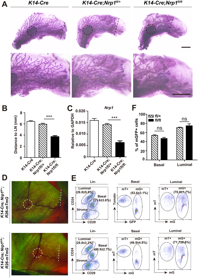
Loss of Nrp1 in basal cells compromises mammary development. (A) Representative images of whole mount carmine staining for 7-week old K14-Cre, K14-Cre;Nrp1 fl/+ and K14-Cre;Nrp1 fl/fl mice. Dash line, forefront of epithelium extension; dash line circle, lymph node (L.N.). Scale bar, top panels, 2mm; bottom panels, 2mm. (B) Quantification of the mammary duct extension in K14-Cre, K14-Cre;Nrp1 fl/+ and K14-Cre;Nrp1 fl/fl mice. The distance from L.N. to epithelium forefront was measured. n = 3 mice in each group. Data are presented as mean ± SEM. ***P < 0.0001. (C) qPCR analysis validating the Nrp1 knockout efficiency using FACS-isolated basal cells. (D) Whole mount imaging of mammary glands with lineage-traced mGFP at 6-week old. (E,F) FACS analysis indicating the proportion of basal and luminal cells, and the percentages of mG+ cells in basal and luminal compartments (E). Quantification indicating no significant differences of mG+ cell percentage in either basal or luminal compartment when comparing the Nrp1 knockout (fl/fl) and the Ctrl (fl/+) (F). n = 3 mice in each group.

