Figure 2.
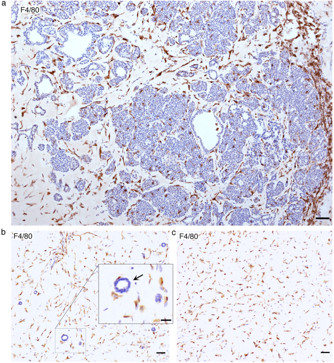
Analysis of stromal infiltrate in transplanted PyMT plugs. (a) Representative image macrophage staining (F4/80) at 2-week showing a transition from ductal- to a tumour-like growth with an increasing concentration of macrophages. Scale bar: 50 µm. (b) Representative image macrophage staining (F4/80) of a primary normal mammary cell transplant at 1-week. Scale bar: 50 µm. Insert close up of a cross-section of a normal duct and surrounding F4/80 positive macrophages, Scale bar: 25 µm. (c) Representative image macrophage staining (F4/80) of an empty Matrigel plug 1-week post-transplantation. Scale bar: 50 µm.

