Figure 1.
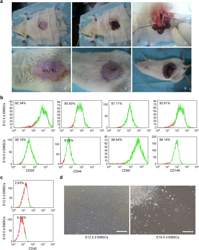
The isolation and identification of rat ectomesenchymal stem cells (EMSCs). (a–d) We isolated EMSCs from embryonic facial processes on day 12.5 and day 19.5 of the embryonic development process though an abdominal operation on the same pregnant SD rat. (a) The abdominal surgery steps are shown in pictures 1–6 The surgery steps in 1–4 were completed on embryonic day 12.5 (E12.5d), and those in 5–6 were completed on embryonic day 19.5 (E19.5d) on the same rat. (1) The SD pregnant rat was intraperitoneally injected with 2.5% phenobarbital sodium (1 ml/100 g) on day 12.5 of embryonic development and was then placed on a rat plate with towels for hair removal and disinfection. (2) Subsequently, the abdomen skin was cut along the abdominal midline, the abdominal muscles were separated, the abdominal cavity was exposed and the embryos were removed from the abdominal cavity. (3) The proximal, distal and mesenteric vessels of the embryos were ligated and the embryos were isolated. (4) Erythromycin (1 ml) and levofloxacin (1 ml) were dropped into the abdominal cavity and the muscle and skin were hierarchically sutured. (5) The same SD pregnant rat continued to grow to day 19.5 of embryonic development; by then, the incision had healed well. (6) The SD pregnant rat was then intraperitoneally injected with 2.5% phenobarbital sodium and was executed to obtain the remaining embryos. (b) The mesenchymal stem cell surface markers (CD29, CD44, CD90 and CD146) and (c) the haematopoietic marker CD45 were detected on EMSCs by the Flow cytometry. (d) The morphologies of the EMSCs separated on E12.5d and E19.5d were observed by optical microscopy. The scale bar represents 50 μm. E12.5d EMSCs, ectomesenchymal stem cells separated at E12.5d and E19.5d EMSCs, ectomesenchymal stem cells separated at E19.5d.

