
Fig. 3.
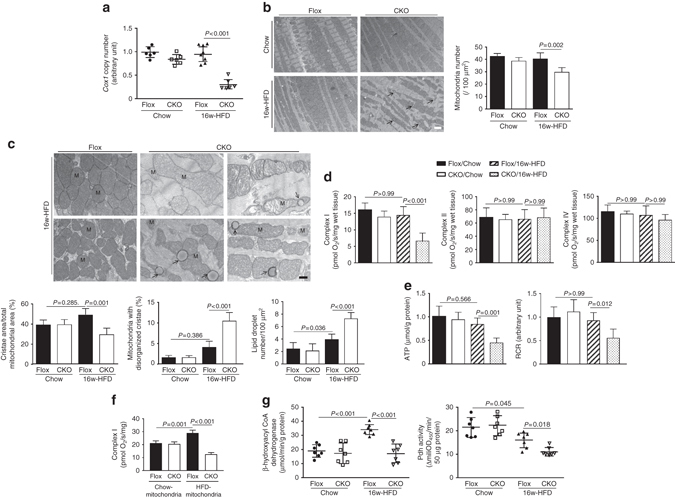
Mitochondrial morphological and functional aberrations in HFD-CKO hearts. a The content of mtDNA was measured by the ratio of Cox1 to cyclophilin A. b Mitochondria was examined by TEM (×1400). Representative images showing reduced and disorganized mitochondria cluttering in the cytosol of HFD-CKO hearts (scale bar: 2 µm). The quantitative analysis of mitochondria number is provided in bar graphs. c Higher magnification (×4800) of TEM images show disarrayed cristae, vacuoles and a reduced electron density of the matrix in the HFD-CKO mitochondria (scale bar: 0.5 µm), M symbolize mitochondria, arrows indicate lipid droplets. Quantitative analysis of cristae area, disorganized cristae in mitochondria, lipid droplet number, respectively, is provided in the bar graphs (n = 6 mice per group). d Respiratory rates of mitochondrial ETC complexes were measured in saponin-permeabilized myocardial fibers. With glutamate/malate as substrates, complex I respiratory rate was found to be decreased in HFD-CKO hearts. e Significantly reduced ATP production in HFD-CKO hearts was determined. Consistent with this, the respiratory control ratio (RCR) was lower in HFD-CKO hearts. f Analysis of complex I in mitochondria homogenates using palmitoylcarnitine as a substrate showed that ERK5-deficient mitochondria of HFD-fed mice had a reduction in FA-driven respiration. g Mitochondrial fuel oxidation was suppressed in HFD-CKO hearts evidenced by activity measurement of β-hydroxylacyl CoA dehydrogenase and pyruvate dehydrogenase (Pdh) (n = 7 mice per group). Data are presented as means ± SD