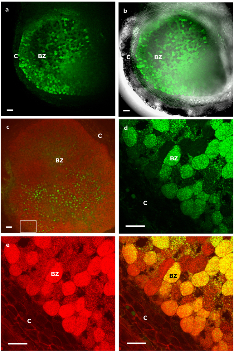
Figure 1.
Longitudinal nodule sections of Lupinus albus coinoculated with Bradyrhizobium sp. CAR08 and Micromonospora ML01-gfp (21 dpi). (a) Green fluorescence signal captured by CLSM of infected cells containing Micromonospora ML01-gfp. (b) Overlay of light and fluorescence images of the nodule section. (c) Green fluorescence localization of ML01-gfp in a nodule section stained with propidium iodide and viewed by CLSM. (d) Higher magnification image captured with the green channel. (e) Higher magnification image captured with the red channel. (f) Composite image of both channels. The white rectangle in image c shows the area where images d-f were captured. C, cortex; BZ, bacteroid zone; dpi, days post inoculation. Bars: 100 µm (a, b, c); 40 µm (d, e, f).

