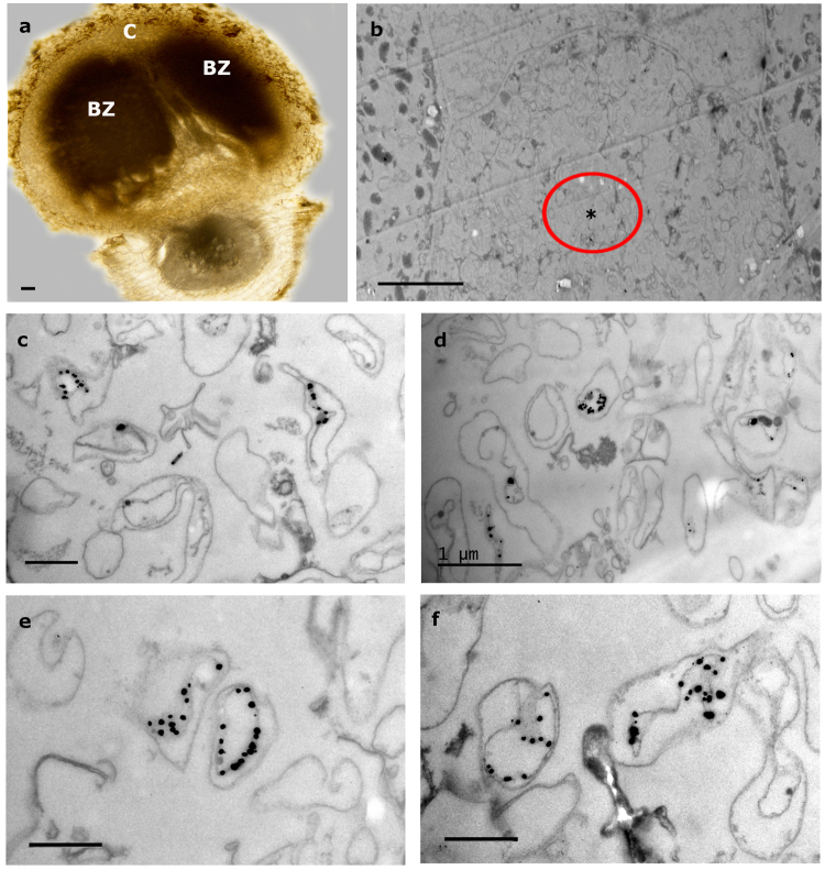
Figure 3.
Immunoelectron microscopic images of lupine nodules infected with Bradyrhizobium CAR08 and Micromonospora ML01-gfp (21 dpi). (a) Light micrograph of a longitudinal nodule section. (b) Detail of an “empty” cell between two infected cells that contain bacteroids. (c–f) Labeled Micromonospora cells found in the area marked with an encircled asterisk in 3b. Bars: (a) 100 µm; (b) 10 µm; (d) 1 µm; 500 nm (c, e, f). C, cortex; BZ, bacteroid zone; dpi, days post inoculation.

