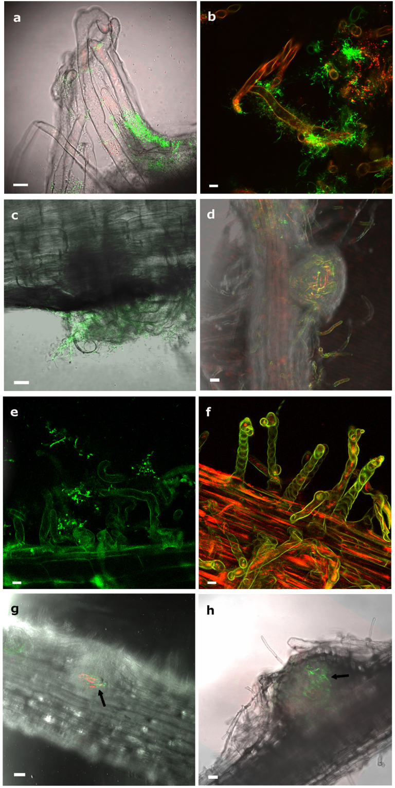
Figure 5.

Infection and colonization of Trifolium and Medicago by Micromonospora ML01-gfp co-inoculated with strains Rhizobium sp. E11-mCh and Sinorhizobium sp. Rm1021-mCh respectively, and observed by CLSM. (a) Trifolium root tip deformations observed 3 dpi and surrounded by Micromonospora ML01-gfp. (b) Micromonospora and Rhizobium sp. co-localized on the root hairs. (c) Nodule primordium and deformed root hairs observed in Trifolium 5 dpi. (d) Young Trifolium nodule observed 11 dpi. The fluorescent bacteria are visible within the internal tissues of the nodule. (e) Attachment of Micromonospora to Medicago root tips showing deformations 6 dpi. (f) Medicago root hair tips forming spiral shapes 12 dpi. (g,h) Medicago nodules at 11 and 13 dpi with green and red fluorescence signals showing strings of bacteria. Because of the thickness of the tissue, the nodules themselves are slightly out of focus. For details see text. Bars: 8 µm (a, b); 10 µm (c, e, f); 75 µm (g, h). dpi, days post infection.
