Figure 3.
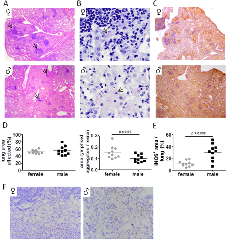
Histological characteristics of Mtb infected lungs. Male and female C57BL/6 were infected via aerosol with Mtb H37Rv and histologically evaluated after 152 days. (A) Granulomatous lesions in H&E stained lung sections (arrows: lymphoid aggregates). (B) Acid fast bacilli (arrows). (C) Immunohistochemical staining of iNOS. Representative micrographs from one mouse out of 10 mice per group are shown (original magnification × 40 in A and C; × 400 in B). (D) Quantitative assessment of total lung area affected by lesions (left graph) and the ratio of lymphoid aggregates to lesion area (right graph). (E) Quantitative evaluation of iNOS expression shown in (C. F) Immunohistochemical staining of neutrophils (Ly6B.2). Representative micrographs from one mouse out of 10 mice per group are shown (original magnification × 100).

