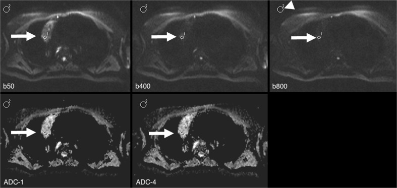
Fig. 3.
A 47-year-old female diagnosed with Hodgkin’s lymphoma. Protocol was identical to that in Fig. 1. In this case, the corresponding region of interest (ROI) 1 (arrow) was placed within the tumour (noise ROI: arrowhead). Apparent diffusion coefficient (ADC) values ranged from 1.800 (ADC_1) to 1.972 (ADC_2; ∆ = 9 %). Note the low remaining signal at b800 (signal-to-noise ratio (SNR) of 3.5) in this heterogeneous lesion

