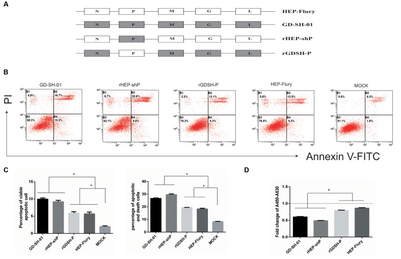
FIGURE 2.
P gene of GD-SH-01 contributes to the enhanced apoptosis induced by wt GD-SH-01. (A) Schematic representations of the gene order of GD-SH-01, HEP-Flury, rHEP-shP, and rGDSH-P. (B) NA cells were infected with GD-SH-01, HEP-Flury, rHEP-shP, or rGDSH-P. Cells in early stage apoptosis are Annexin V+/PI-, while dead cells correspond to those marked Annexin V+. (C) Chart plot of host cell apoptosis at 2 dpi. (D) NA cells were infected with GD-SH-01, HEP-Flury, rHEP-shP, or rGDSH-P, and the cell viability was determined. Data are represented as means ± SD of three independent experiments. One-way ANOVA followed by Bonferroni’s multiple comparison test. ∗p < 0.05.

