Figure 2.
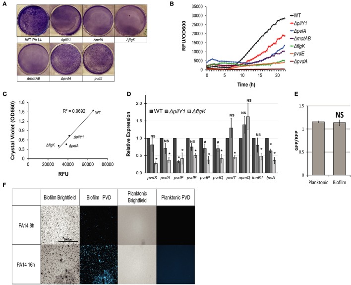
Biofilm formation is necessary for pyoverdine production. (A) Biofilm matrix of PA14 or 6 PA14 mutants in six-well plates stained with 0.1% crystal violet solution. (B) Pyoverdine fluorescence normalized to bacterial growth, measured kinetically over 24 h in biofilm mutants. (C) Scatterplot of pyoverdine and biofilm produced in PA14 biofilm mutants grown in 6-well plate cultures. Biofilm was quantified by resuspending crystal violet stain in 30% acetic acid solution and measuring absorbance at 550 nm. (D) qRT-PCR of genes involved in pyoverdine biosynthesis, secretion, and signaling in WT P. aeruginosa PA14, ΔpilY1 mutant, and ΔflgK mutant. Gene expressions in biofilm mutants are normalized to those in wild-type. (E) Pyoverdine expression in planktonic and biofilm cells after 16 h growth measured by GFP fluorescence from PpvdA-gfp construct normalized to constitutively expressed dsRed fluorescence. (F) Pyoverdine production in PA14 biofilm matrix and planktonic cells imaged using pyoverdine-specific fluorescence filter. Data presented in (A–C), and (F) are representative results from three biological replicates. Error bars in (D,E) represent SEM between three biological replicates. NS corresponds to p > 0.05, # corresponds to p < 0.05, and * corresponds to p < 0.01 (based on Student's t-test). Pyoverdine production curves without bacterial growth normalization are available in Figure S6.

