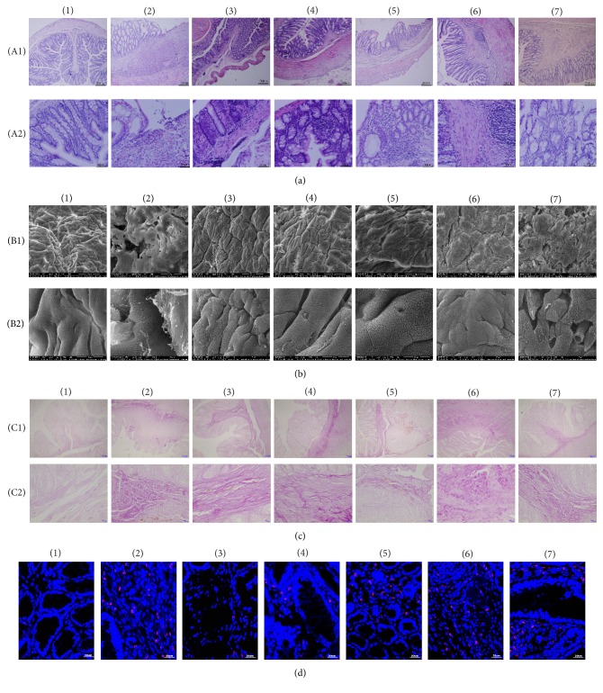
Figure 2.
Effects of Garidisan on the degree of maturity of regenerated mucosa. (a) Histological examination of colon tissue of experimental animals (H&E), (A1) representing 100x magnification and (A2) representing 400x magnification; (b) colonic mucosal SEM of experimental animals, (B1) representing 800x magnification and (B2) representing 5,000x magnification; (c) collagen fiber staining (VG staining) in colons of experimental animals, (C1) representing 100x magnification and (C2) representing 400x magnification. Collagen fibers in panel (c) stained purple; (d) ICAM-1 expression in colonic mucosa and submucosal epithelial of experimental animals. Blue DAPI staining represents nuclear staining, and red represents ICAM-1 expression in cy3 staining. Note that (1) represents normal controls; (2) represents the UC model group; (3) represents the low-dose Garidisan group; (4) represents the moderate-dose Garidisan group; (5) represents the high-dose Garidisan group; (6) represents the SAPA group; and (7) represents the Bupiyichangwan group.

