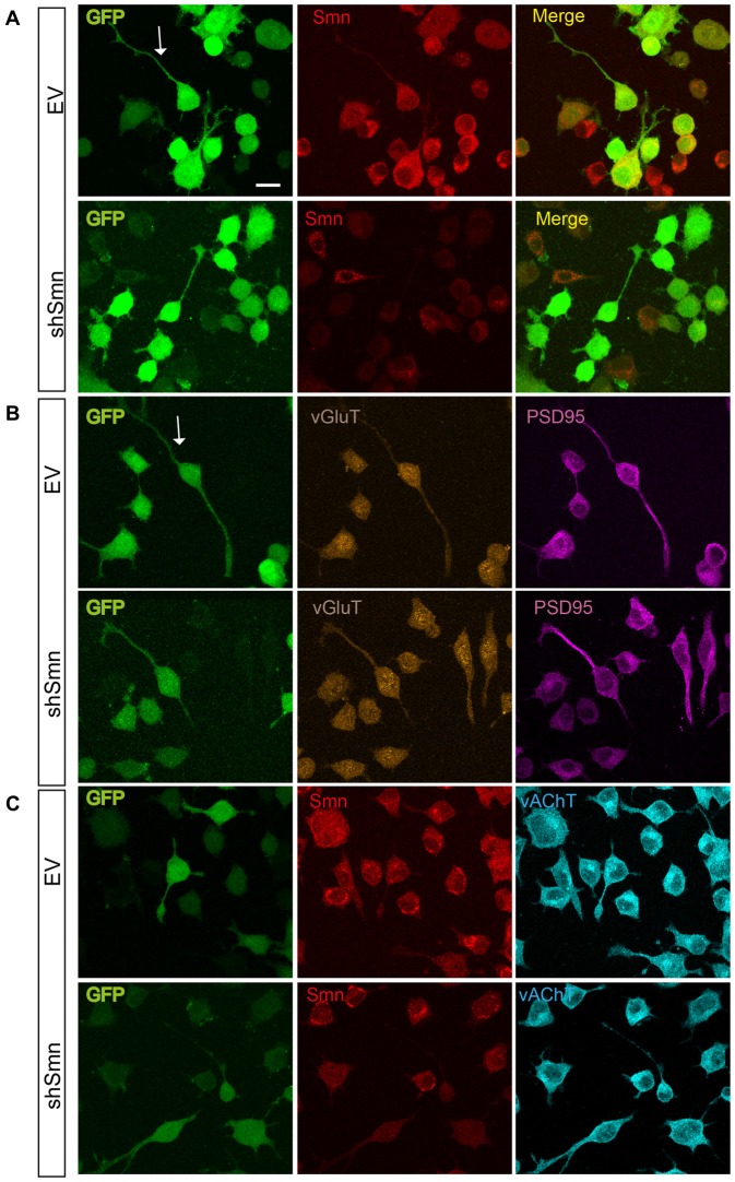
Figure 4.
EV- and shSmn transduced NSC-34 neurons. Confocal images of fixed and immunostained control (EV) and shSmn cultures at DIV3 showing cells with bipolar or pyramidal somata, relatively long neurites and growth cone-like structures (examples marked by arrows). Lentivirally transduced cells display direct green fluorescence (GFP). (A) Representative examples of Smn expression as assessed by immunostaining in EV and shSmn-transduced cells (central panel). (B,C) Immunoreactivity to vGlut (B, central panels), PSD95 (B, right panels), and vesicular acetylcholine transporter (vAChT) (C, right panel) was homogeneous and revealed the absence of synaptic contacts. Calibration bars: 20 μm.

