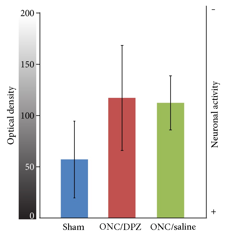
Figure 4.

Cortical neuronal activity measured by thallium autometallography. Optical density (0 (black) to 255 (white) gray levels) was measured within V1 on coronal sections of the brain of rats having been perfused with a thallium solution during monocular flicker visual stimulation (see text). The high gray values correspond to low neuronal activity gradient (scale on the right). The optical density within the visual cortex was not significantly altered 5 weeks after the ONC, as shown for the ONC/DPZ (red) and ONC/saline (green) groups, compared to sham group (blue).
