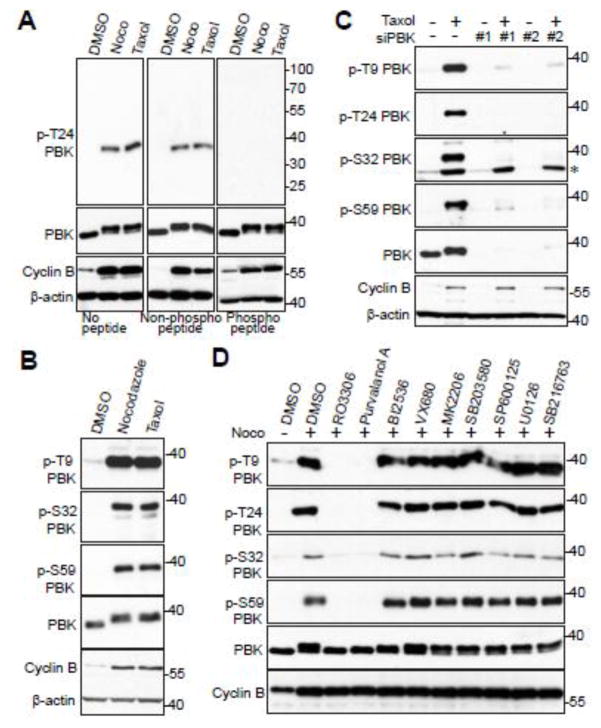
Figure 3. CDK1 phosphorylates PBK in cells.
A, HeLa cells were treated with nocodazole or taxol for 16 hours. The T24 phospho-antibody was incubated for 16 hours at 4°C with either PBS (no peptide), peptide, or phospo-peptide used for immunizing rabbits, before Western blotting. B, HeLa cells were treated with nocodazole or taxol for 16 hours before Western blotting was performed with the indicated antibodies. C, HeLa cells were transfected with scrambled siRNA (control) or siRNA against PBK for 48 h and were further treated with (+) or without (−) taxol for 16 hours. The total cell lysates were subjected to Western blotting with the indicated antibodies. * marks a non-specific band. D, HeLa cells were nocodazole together with or without various kinase inhibitors as indicated. Inhibitors were added with MG132 (to halt cyclin B degradation and prevent mitotic exit) for 1.5 h before harvesting the cells. Total cell lysates were subjected to Western blotting with the indicated antibodies.

