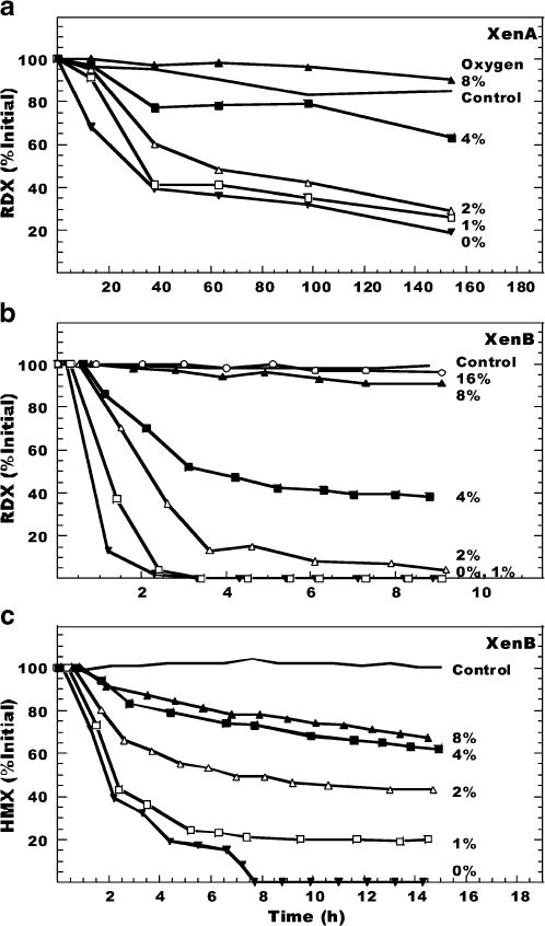
Fig. 3.

Degradation of RDX by purified a XenA and b XenB enzymes and c HMX by purified XenB enzyme under different initial oxygen concentrations. The initial concentrations of RDX and HMX were 55, 83, and 9 μM in a, b, and c, respectively. Control contained ambient oxygen concentration (~20%). Each line represents data from two duplicate vials that were alternatingly sampled during the course of the experiment; therefore, no error bars were calculated. Note difference in x-axis scales
