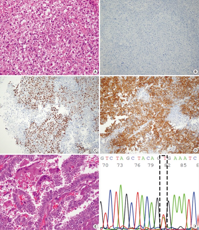
Fig. 2.
Histological and immunohistochemical features of case 2. (A) Metastatic lesion of the brain shows an undifferentiated histology (H & E staining, × 200). (B-D) Metastatic foci of the brain were negative for TTF-1, (B) but positive for PAX8, (C) and BRAF (D) (× 100). (E, F) The neck node, which was removed one year earlier, shows the presence of papillary carcinoma (E) (H & E, × 200) and a missense mutation in the BRAF gene (c.1799T>A) (F).
H & E = hematoxylin and eosin, TTF-1 = thyroid transcription factor-1, PAX8 = paired box gene 8.

