Figure 1.

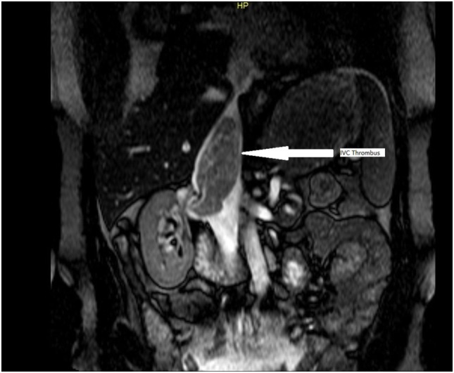
Magnetic resonance imaging image showing extension of the thrombus from the right kidney into the inferior vena cava (IVC) up to the level of the diaphragm, arrow showing IVC thrombus.

Magnetic resonance imaging image showing extension of the thrombus from the right kidney into the inferior vena cava (IVC) up to the level of the diaphragm, arrow showing IVC thrombus.