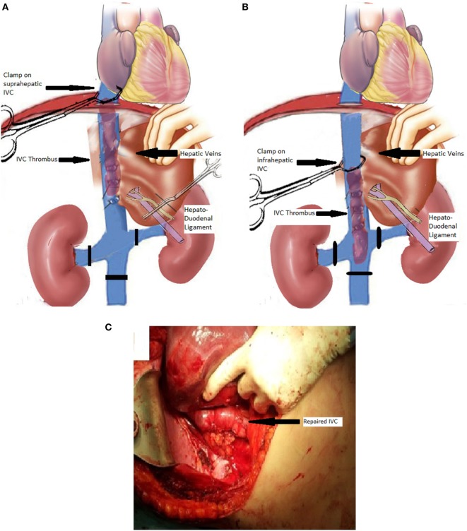
Figure 2.
Schematic view of the procedure in a patient with type II involvement of the inferior vena cava (IVC) with thrombus extending from a right-kidney carcinoma. (A) Schematic view of the IVC, renal veins, liver, hepatic veins, and hepatoduodenal ligament (portal triad structures). The infrarenal IVC and the left renal vein have been encircled with vessel loops (black lines) to allow for subsequent vascular control. A vascular clamp has been placed on the suprahepatic IVC, and the hepatoduodenal ligament has been clamped. (B) The suprahepatic IVC clamp has been exchanged with an infrahepatic IVC clamp after milking the thrombus downwards, and the hepatoduodenal ligament clamping (Pringle maneuver) has been released for hemodynamic stability. (C) Full view of the repaired retrohepatic IVC at the end of the procedure (arrow showing the repaired IVC).

