FIGURE 4.
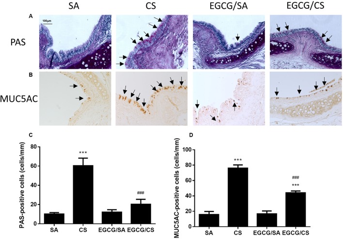
Effect of EGCG on airway mucus secretion in rats. Rats treated with EGCG or control vehicle were killed on day 57 and the left lung was formalin-fixed and sectioned for histology/immunohistochemistry (magnification ×200). (A) Representative photomicrographs of lung sections stained with periodic acid Schiff (PAS). Goblet cells appear as purple staining (arrows) over epithelium. PAS staining revealed increased goblet cell metaplasia after CS exposure and EGCG reduced the CS-induced goblet cell metaplasia. (B) Immunohistochemistry for MUC5AC was performed using an anti-MUC5AC peptide mouse polyclonal antibody, and detected with an anti-mouse/rabbit IgG peroxidase antibody and diaminobenzidine (DAB). MUC5AC-positive staining showed similar phenomenon of mucus secretion of PAS-staining in different groups. Scale bar, 100 μm. Arrows indicate representative cells with positive staining. (C) Quantification of PAS-positive cells per length of epithelium for goblet cells of different groups. (D) Quantification of MUC5AC-positive cells per length of epithelium for mucin of different groups. The results are expressed as means ± SEM; n = 5–6 each group. SA group, water/sham air; CS group, water/cigarette smoke; EGCG/SA group, EGCG (50 mg/kg)/sham air; EGCG/CS, EGCG (50 mg/kg)/cigarette smoke. ∗∗∗p < 0.001 for the comparison to SA group, ###p < 0.001 for the comparison between EGCG/CS and CS group.

