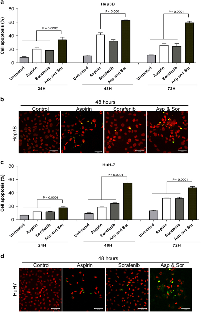
Figure 2.
The synergistic inhibition effects of combination aspirin and sorafenib in HCC cells. Aspirin- and sorafenib-induced apoptosis and DNA fragmentation in Hep3B and HuH-7 cells, respectively. (a and c) Quantification analysis of total cell death from flow cytometry. Annexin V-PE/7-AAD analysis of Hep3B (a) and HuH-7 (c) cells after treated for 24, 48 and 72 h with study agents. Floating and adherent cells were collected after treatment and analyzed by flow cytometry. Points, mean; bars, S.E. (n=3). (b and d) TUNEL staining of Hep3B (b) and HuH-7 (d) cells after 48 h of treatment with study agents. The bright yellow fluorescence spots indicated the apoptotic cells. Nuclei were counterstained with 7-AAD. TUNEL-stained cells were observed at ×40 magnification and scale bars indicate 50 μm.

