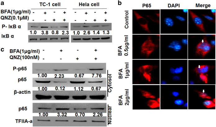
Figure 5.
BFA triggers NF-κB pathway activity in TC-1 tumor cells or HeLa cells. (a) Western blot analysis of p-IðBa and IðBa in whole-cell lysates from TC-1 cells and HeLa cells, which were treated with BFA for 24 h in the presence or absence of QNZ. (b) Immunofluorescence images of TC-1 tumor cells treated with BFA. Arrows point to nuclear transduction of P65. Scale bar: 10 μm. (c) TC-1 cells treated with BFA for 24 h in the presence or absence of QNZ. Alternatively, lysates were subjected to subcellular fractionation to evaluate the presence of p65 in the cytoplasm versus the nucleus, using β-actin and TFIIA-a as control equal loading of cytoplasmic and nuclear fractions, respectively. Protein levels were determined by western blotting compared with those of β-actin.

