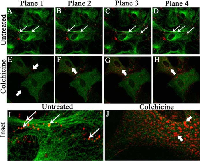
Figure 1.

Localization of Cx43 in primary astrocytes upon colchicine treatment. Primary astrocytes were treated with 100 μm colchicine for 24 h. Control cells were maintained in parallel. Cells were subjected to double-label immunofluorescence with rabbit anti-Cx43 antiserum (red) and mouse anti-β-tubulin antiserum (green). Z-stacking was obtained by a confocal microscope from the base of the cells, at the coverslip (Plane 1), to the medial part of the cells (Plane 4), to observe the distribution of Cx43 with microtubule morphology. Untreated cells showed the presence of Cx43 at both the basal (A, thin arrow) and medial parts of cells (B–D). In contrast, at the basal stack of colchicine-treated cells (E, thick arrow), the presence of Cx43 was minimal. The medial stacks showed the presence of Cx43 mostly inside the cells, showing that Cx43 delivery was restricted upon MT disruption, which was confirmed by disrupted β-tubulin staining (F–H, thick arrow). Digitally magnified insets showed that Cx43 was present on MT threads in a single focal plane, and colocalization was evident, specifically where intensities of Cx43 and β-tubulin were similar (I, ringlike yellow spots, thin arrow). Colchicine-treated cells showed smearlike disrupted β-tubulin signal, whereas Cx43 surface localization was restricted (J, thick arrow).
