Figure 11.
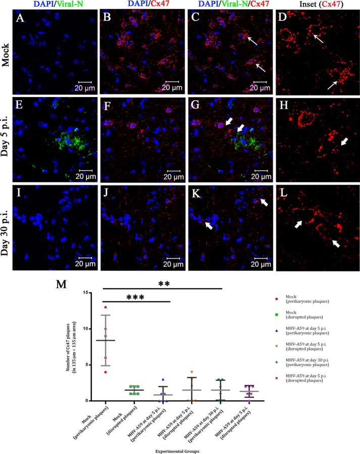
In situ immunofluorescence data on infected brain tissue demonstrated sustained loss of perikaryonic Cx47 signal in MHV-A59–infected mouse brain. Cryosections were obtained from mock– and MHV-A59–infected mouse brains at days 5 and 30 p.i., and double-label immunofluorescence was performed for viral N (green) and Cx47 (red). Nuclei were stained with DAPI (blue). No virus-specific staining was observed for mock-infected brains (A and C), and prominent Cx47 staining was observed around oligodendrocytic somata (B and C (thin arrow); merged image). The characteristic perikaryonic signal of Cx47 was evident (D, inset). At day 5 p.i., MHV-A59–infected brains showed the presence of viral N signal (E and G). Loss of Cx47 signal was observed specifically around the virus-infected area of the brain (F and G (thick arrow)). The inset shows Cx47 immunostaining was disrupted (H, thick arrow). At the peak of demyelination at day 30 p.i., there was no infectious viral particle observed in brain (I and K). In contrast, disrupted Cx47 staining was noticeable in some areas of the brain (J and K (thick arrow)). Normal Cx47-specific signal, visible in oligodendrocytic perikarya, remained depleted at day 30 p.i. (L, inset, thick arrow). Images (with an area of 135 × 135 μm2) obtained from n = 3 biological replicates were quantified for the presence of complete perikaryonic punctate or disrupted signal of Cx47 (M). A reduction of perikaryonic Cx47 plaque count was observed at day 5 as well as at day 30 p.i. At day 5 p.i., ∼7.6 Cx47 plaques were reduced in the MHV-A59 infected mice, in an area of 135 × 135 μm2 (***, p < 0.001; t test). At day 30 p.i., ∼6.9 intact Cx47 plaques were reduced in an area of 135 × 135 μm2 (**, p < 0.01; t test) (M). Error bars, S.D.

