Figure 2.
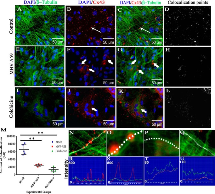
Altered association of Cx43 with microtubule in primary astrocytes upon MHV-A59 infection and colchicine treatment. Primary astrocytes were infected with MHV-A59 at an MOI of 2 or treated with 100 μm colchicine for 24 h. Control cells were maintained in parallel. Cells were subjected to double-label immunofluorescence with rabbit anti-Cx43 antiserum (red) and mouse anti-β-tubulin antiserum (green). Cells were counterstained with DAPI (blue). In control astrocytes, punctate Cx43 (B, thin arrow) was observed to be closely associated with β-tubulin (A), and Cx43 staining was aligned along the typical radial structure of MTs (C, thin arrow). Spots of colocalized signal are shown in D. Upon infection of the cells with MHV-A59, Cx43 was retained in the intracellular compartment (F, thick arrow). Although the MT morphology appeared to be normal (E), intracellular compartment–retained Cx43 had a minimal association with the MT network (G, thick arrow). The intensity and number of colocalization spots were reduced (H). Upon colchicine treatment, the MT network was depolymerized in the primary astrocytes, and diffuse tubulin staining was observed in the cytosol (I). Colchicine treatment showed retention of Cx43 in the intracellular compartment, predominantly in the perinuclear area (J and K, thick arrow). The number of colocalization spots was reduced significantly (L). The number of colocalization points compared between experimental groups showed there was an ∼62.8% reduction in virus-infected cells compared with mock-infected cells (**, p < 0.01; Mann–Whitney U test) and ∼80.3% reduction in colchicine-treated cells compared with mock-infected cells (**, p < 0.01; Mann-Whiney U test). Comparison of all three groups by Kruskal–Wallis ANOVA showed that differences were significant (****, p < 0.0001; M). Five fields were analyzed for each group from n = 3 experiments. Digitally magnified images show that Cx43 molecules were aligned along a MT thread (N and O), whereas intracellular compartment–retained Cx43 did not show such alignment (P and Q). Association of Cx43 molecules on a single MT thread is shown (red line, Cx43; green line, tubulin). Cx43 molecules showed high-intensity peaks on MT threads in mock-infected cells (R and S), but not in virus-infected cells (T and U). Error bars, S.D. AU, arbitrary units.

